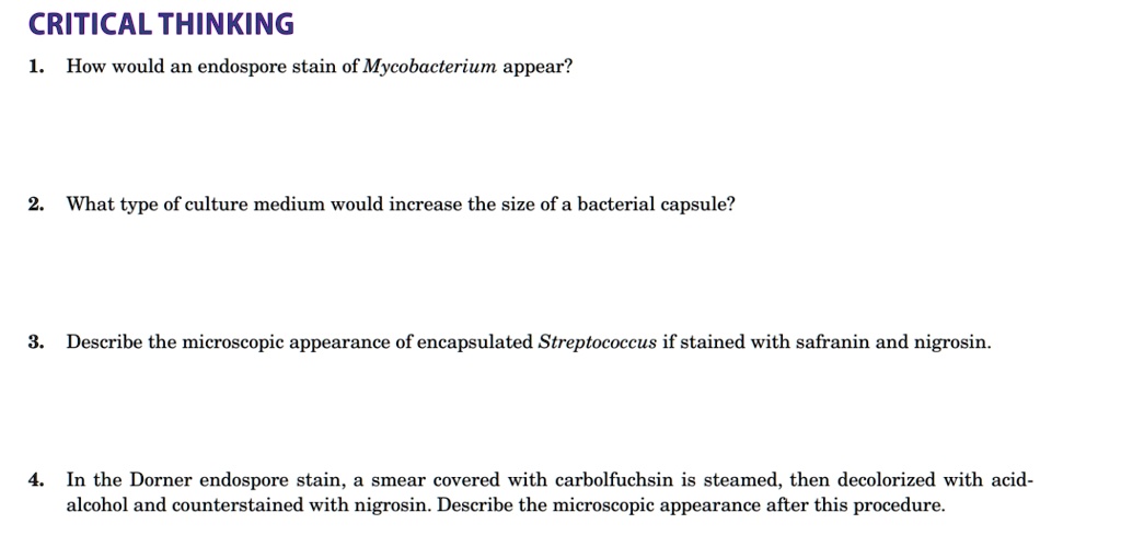
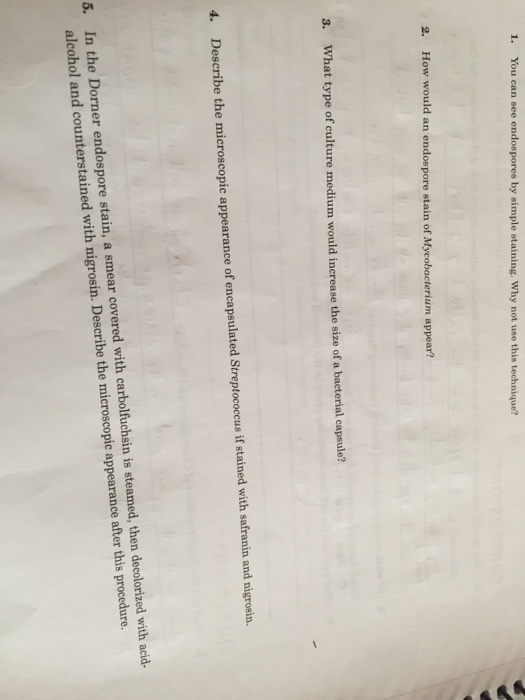
D escribe the difference in appearance between absorption and emission lines. 10 rows Describe the microscopic appearance of encapsulated Streptococcus if stained with safranin and nigrosin The stretococcal cells would be red surrounded by a clear capsule against a black background In the Dorner endospore stain a smear with carbolfuchsin is steamed then decolorized with acid alcohol and counterstained with nigrosin.

Exercise 9 Structural Stains Endospore Capsule Flagella Flashcards Quizlet
As cellular division of Streptococcus spp.

. I f I build a big building with pretty stained glass and offer you salvation of my own accord will you come to my church. Illnesses caused by streptococcus include strep throat strep pneumonia scarlet fever rheumatic fever and rheumatic heart valve damage glomerulonephritis the skin disorder erysipelas and PANDAS. Describe the microscopic appearance of encapsulated Streptococcus if stained with safranin and nigrosin.
Group D grow well at between 10 degree Celsius and 45 degree Celsius. Check out a sample Q. Like many other bacteria Streptococcus bacteria are small in size ranging from 05 to 20 micrometers in diameter.
It is the causative agent of acute pharyngitis impetigo erysipelas necrotizing fasciitis flesh-eating bacteria and myositis. Often dry colonies are observed. S pneumoniae appears as a 05-125 μm diplococcus typically described as lancet-shaped but sometimes difficult to distinguish morphologically from other streptococci.
Describe the microscopic appearance of an encapsulated Escherichiae if stained with nigtosin followed by safranin. Escherichia stained with nigrosine followed by safranin would appear as a red bacteria with a clear capsule and a purple background. The bacteria would be red the capsule clear and the background purplish-black In the Dorner endospore stain a smear covered with Carbolfuchsin is steamed then decolorized with acid-alcohol and counter-stained with nigrosin.
MICROSCOPIC APPEARANCE MACROSCOPIC APPEARANCE Streptococcal colonies vary in color from gray to whitish and usually glisten. Cocci in clusters short chains diplococci and single cocci. Here however the majority of species are less than 2um in size.
Red bacteria clear capsule with purpleblack background. Staphylococci divide in various directions multiple axes. More than 90 different serotypes are known and these types differ in virulence.
It is usually. Streptococcus pyogenes or Group A streptococcus GAS is mostly known for streptococcal sore throat strep throat. Under a microscope streptococcus bacteria look like a twisted bunch of round berries.
In the Dorner endospore stain a smear covered with carbolfuchsin is steamed then decolorized with acid-alcohol and counterstained with nigrosin. Describe the microscopic appearance of an encapsulated Streptococcus if stained with Congo Redacid-alcohol and safranin. Pneumoniae is a fastidious bacterium growing best at 35-37C with 5 CO 2 or in a candle-jar.
Streptococcal cultures older than the logarithmic phase which is the most active growth period of a culture may lose their Gram-positive staining characteristics. Despite the name the organism causes many types of pneumococcal infections other than pneumonia. Pyogenes is the cause of many important human diseases ranging from mild superficial skin infections to life-threatening systemic diseases.
Describe the microscopic appearance of encapsulated Streptococcus if stained with safranin and nigrosin. A chain of round cells. It is an infrequent but usually pathogenic part of the skin flora.
Cultural Characteristics of Streptococcus. In the Dorner endospore stain a smear covered with carbolfuchsin is steamed then decolorized with acid-alcohol and counterstained with nigrosin. Describe the microscope appearance of encapsulated Streptococcus if stained with safranin and nigrosin.
Carbolfuchsinwill leave the endospores red or pink the nigrosin will make the background dark. Identification and Characterization of Streptococcus pneumoniae. They grow best at 37 degree Celsius.
Division occurs in one linear direction single axis. Generally the word microscopic cannot be used to describe a sound. It has a polysaccharide capsule that acts as a virulence factor for the organism.
It is a gram-positive cocci that mostly occurs as chains and occasionally in pairs. They are usually found in pairs diplococci but are also found singly and in short chains. Individual bacteria are between 05 and.
Many variations exist among Lancefield groups. Occurs along a single axis or plane these bacteria grow in pairs or chains. Encapsulated strains may appear mucoid.
Positive Catalase is an enzyme to converts hydrogen peroxide to water and oxygen gas. A group of bacteria that causes a multitude of diseases. Pneumoniae are alpha hemolytic a term describing how the cultured bacteria break down red blood cells for the purpose of classification.
Want to see the full answer. If 5x instead of 10x oculars were used in your microscope with the same objectives. Pneumoniae may occur intracellularly or extracellularly as gram-positive lanceolate diplococci but can also occur as single cocci or in short chains of cocci.
With regards to shape Streptococci may appear spherical or ovoid in shape. They are nonmotile and non-spore forming. Describe the microscope appearance of encapsulated streptococcus if stained with safarin and nigrosin.
These cocci measure between 05 and 2 μm in diameter. The word microscopic is often a dictation of visual size - ie requiring a microscope to. Streptococci are coccoid bacterial cells microscopically and stain purple Gram-positive when Gram staining technique is applied.
METABOLIC PROPERTIES Facultatively or strictly anaerobic. Lanceolate ovoid cocci in short chains diplococci and single cocci. Describe the microscope appearance after this procedure.
Solution for Describe the microscopic appearance of encapsulated Streptococcus if stained with safranin and nigrosin. Growth is poor on solid media or broth. The encapsulated gram-positive coccoid bacteria have a distinctive morphology on gram stain the so-called lancet shape.
Describe the microscopic appearance after this procedure. Then decolorized with acid-alcohol and countersigned with nigrosin. Streptococcus pneumoniae are Gram-positive bacteria in the shape of a slightly pointed cocci.
Describe the microscopic appearance of an encapsulated Escherichiae if stained with nigrosin and basic methylene blue. They are aerobic and facultative anaerobes. Describe the microscope appearance after this procedure.
Start your trial now. Requires a nutritionally rich media for growth. First week only 499.

Staining Variation Between Figs A E And Within Figs E K Download Scientific Diagram

Solved Critical Thinking How Would An Endospore Stain Of Mycobacterium Appear What Type Of Culture Medium Would Increase The Size Of A Bacterial Capsule Describe The Microscopic Appearance Of Encapsulated Streptococcus If Stained

Solved Critical Thinking 1 How Would An Endospore Stain Of Chegg Com

Solved How Would An End Spores Again Of Mycobacterium Chegg Com
0 Comments